Macule
| Macule | |
|---|---|
.jpg) | |
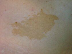
| Cafe-au-lait macule | |
| Specialty | Dermatology |
| Symptoms | |
| Types | Solitary, multiple[1] |
| Diagnostic method | Appearance[2] |
| Differential diagnosis | Several includiong: freckles, lentigo, petechiae, junctional nevus, idiopathic guttate hypomelanosis, viral exanthems[3] |
A macule is a small flat skin spot that is neither raised nor depressed.[1] It may be an early stage of another skin sign, and occurs in varied sizes, shapes and colours.[1][4] Its border may be sharp or blurred and when touched, it feels like the surrounding normal skin.[1] Colors vary from dark brown/black, light, red, purplish, and pink.[3] It's size is generally less than 2cm in diameter, though when merged form a larger appearing patch.[2] Shapes vary from round, to oval, and irregular.[3]
Diagnosis is generally made by its appearance; the color, size and number of the macules.[5] Freckles, lentigo, petechiae and junctional nevus are macules.[3] Idiopathic guttate hypomelanosis and viral exanthems present with maculles.[3] Other conditiions in which macules are a feature include oral melanotic macule and focal argyrosis.[5]
Definition

A macule is a small flat skin spot, that is neither raised or depressed.[1] The term is derived from the latin for stain and describes a color change.[2]
Signs and symptoms
It may be an early stage of another skin sign, and occurs in varied sizes, shapes and colours.[1][4] Its border may be sharp or blurred and when touched, it feels like the surrounding normal skin.[1] It may be dark brown/black, light, or red.[2] It is generally less than 2cm in diameter; larger ones are termed a patch.[2]
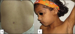
Solitary macules
-
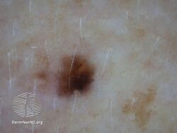
Mole - solitary macule
-
-
.jpg)
.jpg)
.jpg)
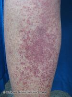
Multiple pink/red/purplish macules
-
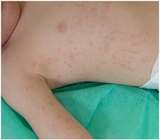
Salmon-macular rash (systemic juvenile idiopathic arthritis) - many red macules
-
Chicken pox - macules with papules
-
Petechiae
-
Exanthem

.jpg)
.jpg)
.jpg)
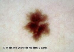
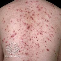
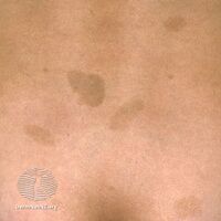
Multiple dark macules
-
Multiple macules - many brown macules
-
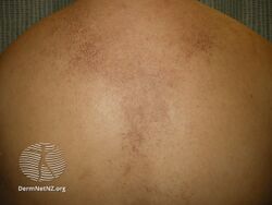
Macular amyloidosis
-

Freckles
-

.jpg)

.jpg)
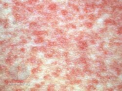
Multiple light macules
-
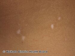
Vitiligo macules - many light macules
-
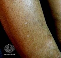
Idiopathic guttate hypomelanosis
-
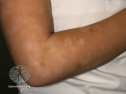
Pityriasis alba: light macules that merge
.jpg)
.jpg)
.jpg)
Diagnosis
Diagnosis is generally made by its appearance; the color, size and number of the macules.[5] Macules are a feature of conditions including oral melanotic macule, ephesus and focal argyrosis.[5]
Differential diagnosis
Common macule include drug reaction, junctional nevus, viral rash, freckles, and chloasma[6]
See also
Reference
- ↑ 1.0 1.1 1.2 1.3 1.4 1.5 1.6 1.7 James, William D.; Elston, Dirk; Treat, James R.; Rosenbach, Misha A.; Neuhaus, Isaac (2020). "2. Cutaneous signs and diagnosis". Andrews' Diseases of the Skin: Clinical Dermatology (13th ed.). Edinburgh: Elsevier. p. 11. ISBN 978-0-323-54753-6.
- ↑ 2.0 2.1 2.2 2.3 2.4 2.5 Morris-Jones, Rachael (2019). "1. Introduction". In Morris-Jones, Rachael (ed.). ABC of Dermatology (7th ed.). Hoboken: Wiley Blackwell. pp. 3–4. ISBN 978-1-119-48899-6. Archived from the original on 2023-02-09. Retrieved 2023-02-09.
- ↑ 3.0 3.1 3.2 3.3 3.4 3.5 Bolognia, Jean L.; Schaffer, Julie V.; Duncan, Karynne O.; Ko, Christine (2022). "1. Basics". Dermatology Essentials (2nd ed.). Elsevier. p. 4. ISBN 978-0-323-70971-2. Archived from the original on 2024-01-03. Retrieved 2024-01-02.
- ↑ 4.0 4.1 Oakley, Amanda. "Terminology in dermatology". dermnetnz.org. Archived from the original on 9 March 2022. Retrieved 16 April 2022.
- ↑ 5.0 5.1 5.2 5.3 Langlais, Robert P.; Miller, Craig S.; Gehrig, Jill S. (2017). "18. Diagnostic and descritive terminology: macule, patch, erosion, ulcer". Color Atlas of Common Oral Diseases, Enhanced Edition (5th ed.). Burlington: Jones & Bartlett Learning. p. 18. ISBN 978-1-284-24098-6. Archived from the original on 2023-02-09. Retrieved 2023-02-09.
- ↑ Hopcroft, Keith; Forte, Vincent (2020). "Skin: macules". Symptom Sorter. Boca Raton: CRC Press. pp. 405–407. ISBN 978-0-367-46810-1. Archived from the original on 2023-09-19. Retrieved 2023-09-19.