Venous lake
| Venous lake | |
|---|---|
| Other names: Phlebectasis[1] | |
 | |
| Venous lake of the lip | |
| Specialty | Dermatology |
| Symptoms | Small dark blue compressible bump in skin[1] |
| Usual onset | Elderly |
| Diagnostic method | Visualisation[1] |
| Treatment | Light electrocautery, laser ablation, infrared coagulation, radiofrequency ablation, injecting it with 1% polidocanol, cryotherapy[1] |
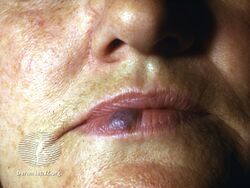
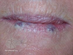
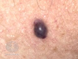
A venous lake is a small dark blue compressible bump in the skin.[1] There is typically just one of them and it is commonly found on sun-exposed surfaces of the lip border, face and ears.[2] It tends to feel soft and can measure 0.2- to 1-cm.[2] Lesions generally occur among the elderly.[3]
The blue color is of blood.[1] Though these lesions may resemble nodular melanoma, the lack of induration, slow growth, and lightening appearance upon diascopy suggest against it, and indicate a vascular lesion.[4] Additionally, lack of pulsation distinguishes this lesion of the lower lip from a tortuous segment of the inferior labial artery.[5]
Treatment options include light electrocautery, laser ablation, infrared coagulation, radiofrequency ablation injecting it with 1% polidocanol, and cryotherapy.[1]
The term was coined by American physician William Bennett Bean in 1956.[6]
Cause
The cause is unknown; however it is thought to be associated with sun exposure, leading to a dilated blood-filled vascular channel[2] "...lined with a singled layer of flattened endothelial cells and a thin wall of fibrous tissue filled with red blood cells."[4]
Treatment
Treatment options include light electrocautery, laser ablation, infrared coagulation, radiofrequency ablation, injecting it with 1% polidocanol, and cryotherapy.[1]
Images
-

Venous lake of the ear
-

Venous lake of the ear
-
Venous lake lower lip
-
Venous lake lower lip
-
Venous lake


.jpg)
.jpg)
.jpg)
See also
Footnotes
- ↑ 1.0 1.1 1.2 1.3 1.4 1.5 1.6 1.7 James, William D.; Elston, Dirk; Treat, James R.; Rosenbach, Misha A.; Neuhaus, Isaac (2020). "28. Dermal and subcutaneous tumors: venous lake". Andrews' Diseases of the Skin: Clinical Dermatology (13th ed.). Edinburgh: Elsevier. pp. 603–604. ISBN 978-0-323-54753-6. Archived from the original on 2023-04-29. Retrieved 2023-04-29.
- ↑ 2.0 2.1 2.2 Habif, Thomas P. Clinical Dermatology: A Color Guide to Diagnosis and Therapy. Mosby, Inc. 2004. Page 825. ISBN 0-323-01319-8.
- ↑ Rapini, Ronald P.; Bolognia, Jean L.; Jorizzo, Joseph L. (2007). Dermatology: 2-Volume Set. St. Louis: Mosby. p. 1620. ISBN 1-4160-2999-0.
- ↑ 4.0 4.1 Wolff and Johnson. Fitzpatrick's Color Atlas and Synopsis of Clinical Dermatology. The McGraw-Hill Companies. 2005. Page 192. ISBN 0-07-144019-4.
- ↑ Sauer, Gordon. Manual of Skin Diseases. Lippincott. 1985. Page 315. ISBN 0-397-50668-6.
- ↑ Mulliken, John B. (2013). "13. Capillary malformations, hyperkeratotic stains, telangiectasias, and miscellaneous vascular blots". In Mulliken, John B.; Burrows, Patricia E.; Fishman, Steven J. (eds.). Mulliken and Young's Vascular Anomalies: Hemangiomas and Malformations (2nd ed.). Oxford University Press. p. 553. ISBN 978-0-19-972254-9. Archived from the original on 2023-07-02. Retrieved 2023-05-19.
External links
| Classification |
|
|---|---|
| External resources |