Kerion
| Kerion | |
|---|---|
 | |
| Kerion | |
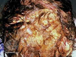
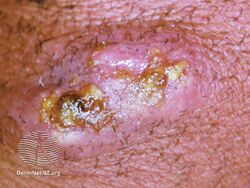
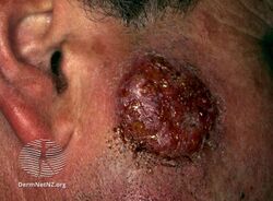
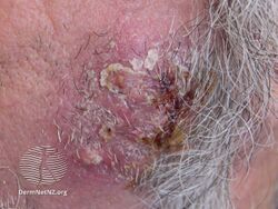
Kerion Celsi is the result of the host's response to a fungal ringworm infection of the hair follicles of the scalp (occasionally the beard) that can be accompanied by secondary bacterial infection(s). It usually appears as raised, spongy lesions, and typically occurs in children.[1] This honeycomb is a painful inflammatory reaction with deep suppurative lesions on the scalp. Follicles may be seen discharging pus. There may be sinus formation and rarely mycetoma-like grains are produced. It is usually caused by dermatophytes (fungal infections of the skin affecting humans and animals) such as Trichophyton verrucosum, T. mentagrophytes,[1] and Microsporum canis. Treatment with oral griseofulvin common.[1]
Signs and symptoms
There is generally a "boggy" raised plaque on the scalp.[2] There may be loss of hair as hair will come out easily.[3] There may be a fever. It can look like impetigo.[4]
-
Kerion
-
Kerion
-
Kerion
-
Kerion
.jpg)
.jpg)
.jpg)
.jpg)
Cause
It is most commonly due to a hypersensitivity reaction to the dermatophyte infection, most commonly T. verrucosum or T. tonsurans.[2]
Diagnosis
The basis for the diagnosis of Kerion is clinical finding, positive microscopic examinations (such as positive KOH preparation, Lactophenol cotton blue wet mount, Chicago sky blue stained (CSB) slide, Calcofluor white stained slide, Periodic acid–Schiff stained slide, and Gomori’s methenamine silver stained slide), mycological culture and modern molecular tests (such as PCR-reverse line blot test, real-time PCR test, multiplex PCR test, PCR-ELISA test, and MALDI-TOF test) of clinical specimens. Wood's lamp (blacklight) examination will reveal a bright green to yellow-green fluorescence of hairs infected by Trichophyton mentagrophytes var. Mentagrophytes, in kerion infection caused by Trichophyton verrucosum Invaded hairs show an ectothrix infection and fluorescence under Wood's ultra-violet light has been noted in cattle but not in humans.[5]
Sometimes, there is growth of organisms.[6]
Treatment
Unlike most other manifestations of Tinea dermatophyte infections, Kerion is not sufficiently treated with topical antifungals and requires systemic therapy. Typical therapy consists of oral antifungals, such as griseofulvin or terbinafine, for a sustained duration of at least 6-8 weeks depending on severity. Successful treatment of kerion often requires empiric bacterial antibiotics given the high prevalence of secondary bacterial infection.[7]
See also
References
- ↑ 1.0 1.1 1.2 Uprety, Shraddha; Sharma, Ramesh (2016-09-08). "Kerion — A Boggy Lump". New England Journal of Medicine. 375 (10): 980. doi:10.1056/NEJMicm1514152. PMID 27602670.
- ↑ 2.0 2.1 Johnstone, Ronald B. (2017). "25. Mycoses and Algal infections". Weedon's Skin Pathology Essentials (2nd ed.). Elsevier. p. 440. ISBN 978-0-7020-6830-0. Archived from the original on 2021-05-25. Retrieved 2021-05-25.
- ↑ "Management of Tinea Capitis". The International Foundation for Dermatology. Archived from the original on March 26, 2018. Retrieved January 21, 2015.
- ↑ "Cause of Kerion Ringworm Scalp Condition, Kerions Treatment". Health Blurbs. Archived from the original on January 10, 2018. Retrieved January 21, 2015.
- ↑ Khalid, Mohamed (2019). "LABORATORY DIAGNOSIS OF THE CAUSATIVE DERMATOPHYTES OF TINEA CAPITIS" (PDF). World Journal of Pharmaceutical Research. 8 (6): 85–99. doi:10.20959/wjpr20196-14850 (inactive 2021-01-11). Archived from the original (PDF) on 2019-05-03. Retrieved 4 May 2019.
{{cite journal}}: CS1 maint: DOI inactive as of January 2021 (link) - ↑ L. C. Fuller; F. J. Child; G. Midgley; E. M. Higgins (March 8, 2003). "Diagnosis and management of scalp ringworm". BMJ. 326 (7388): 539–541. doi:10.1136/bmj.326.7388.539. PMC 1125423. PMID 12623917.
- ↑ "Kerion". DermNet New Zealand. Archived from the original on December 22, 2017. Retrieved December 19, 2017.
External links
| Classification |
|---|