File:Sisteronia.png
Jump to navigation
Jump to search
Size of this preview: 460 × 599 pixels. Other resolutions: 184 × 240 pixels | 369 × 480 pixels | 590 × 768 pixels | 786 × 1,024 pixels | 2,037 × 2,652 pixels.
Original file (2,037 × 2,652 pixels, file size: 4.79 MB, MIME type: image/png)
| Description |
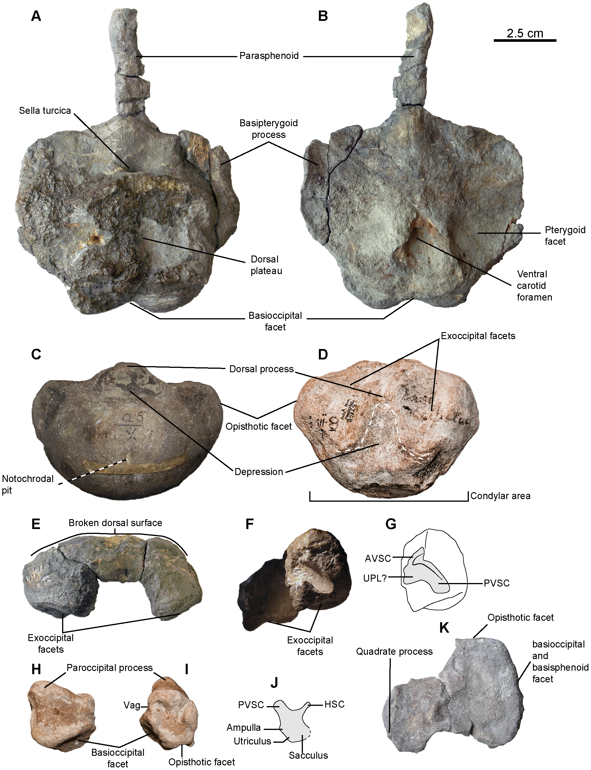
Sisteronia seeleyi, basicranium. A, B: basisphenoid (RGHP SI 2) in dorsal (A) and ventral (B) views. C: basioccipital (CAMSM B57943) in posterior view. D: holotype basioccipital (CAMSM B58257_67) in dorsal view. E–G: supraoccipital (RGHP SI 2) in posterior (E) and anterolateral (otic) (F, G) views. H–J: left opisthotic (CAMSM B58257_67) in posterior (H) and anterior (otic) (I, J) views. K: left stapes (RGHP SI 2) in posterior view. Note the extremely reduced (nearly absent) extracondylar area of the basioccipital, a platypterygiine synapomorphy, and the dorsal process posterior to a triangular depression (delineated by the thick dotted line) on the basioccipital, an autapomorphy of Sisteronia seeleyi. Abbreviations: AVSC: impression of the anterior vertical semicircular canal of the otic labyrinth; HSC: impression of the horizontal semicircular canal of the otic labyrinth; PVSC: impression of the posterior vertical semicircular canal of the otic labyrinth; UPL: impression of the utricular portion of the otic labyrinth; Vag: vagus foramen. |
| Date | |
| Source | http://www.plosone.org/article/info%3Adoi%2F10.1371%2Fjournal.pone.0084709 |
| Author | Valentin Fischer, Nathalie Bardet, Myette Guiomar, Pascal Godefroit |
|
This file is licensed under the Creative Commons Attribution 2.5 Generic license.
|
This file was published in a Public Library of Science journal. Their website states that the content of all PLOS journals is published under the Creative Commons Attribution 4.0 license (or its previous version depending on the publication date), unless indicated otherwise.
|
File history
Click on a date/time to view the file as it appeared at that time.
| Date/Time | Thumbnail | Dimensions | User | Comment | |
|---|---|---|---|---|---|
| current | 18:31, 22 January 2014 | | 2,037 × 2,652 (4.79 MB) | commons>FunkMonk | {{Information |Description=Sisteronia seeleyi, basicranium. A, B: basisphenoid (RGHP SI 2) in dorsal (A) and ventral (B) views. C: basioccipital (CAMSM B57943) in posterior view. D: holotype basioccipital (CAMSM B58257_67) in dorsal view. E–G: supra... |
File usage
There are no pages that use this file.