File:Ribonuclease H structure comparison.png
Jump to navigation
Jump to search
Size of this preview: 530 × 599 pixels. Other resolutions: 212 × 240 pixels | 425 × 480 pixels | 679 × 768 pixels | 906 × 1,024 pixels | 2,300 × 2,600 pixels.
Original file (2,300 × 2,600 pixels, file size: 1.27 MB, MIME type: image/png)
File history
Click on a date/time to view the file as it appeared at that time.
| Date/Time | Thumbnail | Dimensions | User | Comment | |
|---|---|---|---|---|---|
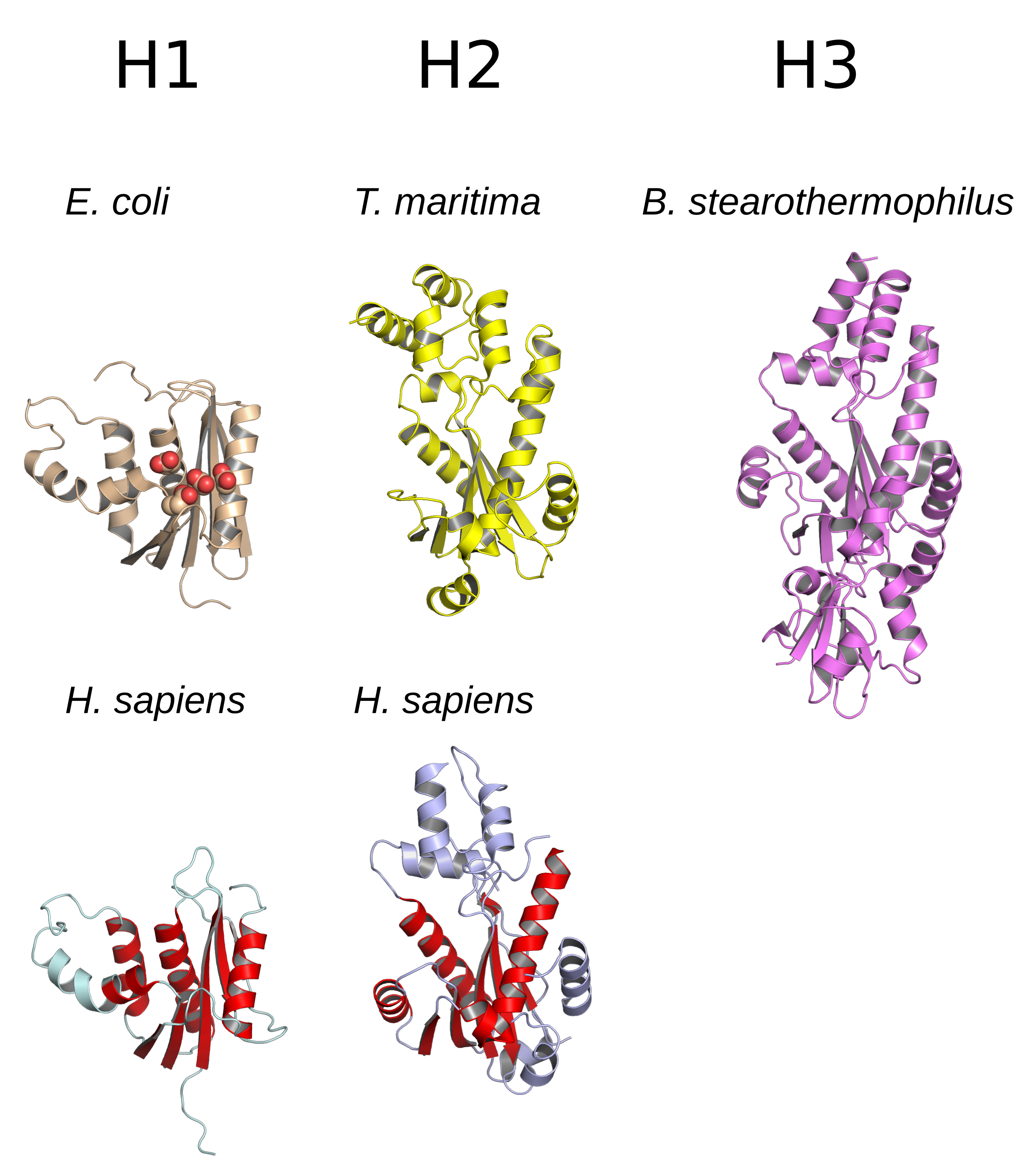
| current | 01:34, 2 March 2017 | | 2,300 × 2,600 (1.27 MB) | commons>Opabinia regalis | {{Information |Description ={{en|1=Comparison of structures of ribonuclease H proteins of types H1, H2, and H3 (HIII in prokaryotic nomenclature). In the E. coli structure, the four conserved active residues are shown as spheres. In the two human st... |
File usage
There are no pages that use this file.