File:Pyomyositis MRI.jpg
Jump to navigation
Jump to search
Size of this preview: 479 × 600 pixels. Other resolutions: 192 × 240 pixels | 383 × 480 pixels | 766 × 959 pixels.
Original file (766 × 959 pixels, file size: 74 KB, MIME type: image/jpeg)
| Description |
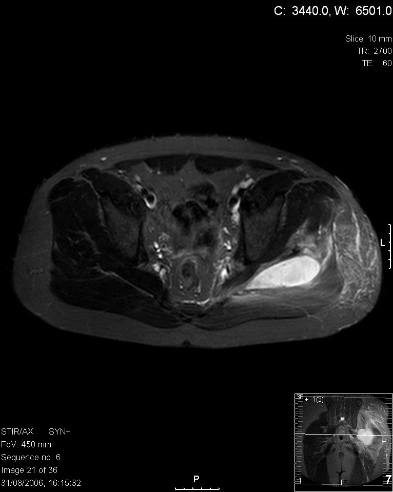
English: Transverse T2 magnetic resonance imaging section through the hip region showing abscess collection in relation to the sciatic nerve in a patient with pyomyositis who presented with sciatica. |
| Date | Published: 12 June 2008 |
| Source | Gluteal pyomyositis in a non-tropical region as a rare cause of sciatic nerve compression: a case report |
| Author | Tamer Kamal, Mathew Hall, Ashraf Moharam, Michael Sharr and Jonathan Walczak |
| Permission (Reusing this file) |
This file is licensed under the Creative Commons Attribution 2.0 Generic license.
|
| Annotations | This image is annotated: View the annotations at Commons |
File history
Click on a date/time to view the file as it appeared at that time.
| Date/Time | Thumbnail | Dimensions | User | Comment | |
|---|---|---|---|---|---|
| current | 20:15, 26 June 2008 | | 766 × 959 (74 KB) | commons>Stevenfruitsmaak | {{Information |Description={{en|1=Transverse T2 magnetic resonance imaging section through the hip region showing abscess collection in relation to the sciatic nerve in a patient with pyomyositis who presented with sciatica.}} |Source=[http://www.jmedical |
File usage
There are no pages that use this file.