File:Pulmonary-mucormycosis-1.jpeg
Original file (597 × 630 pixels, file size: 59 KB, MIME type: image/jpeg)
Summary
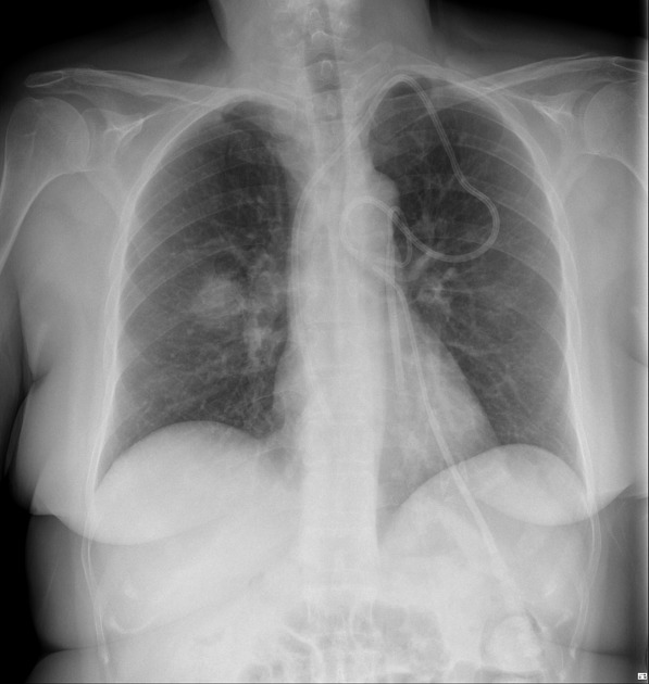
Author:Case courtesy of Dr David Holcdorf, Radiopaedia.org, rID: 64718
Source:https://radiopaedia.org/cases/pulmonary-mucormycosis-1?lang=gb
Description:35 year old febrile female who is immunosuppressed. New 3 cm nodule/mass in the apical segment right lower lobe. Findings are concerning for infection in this setting, and given the patient's immunosuppressed state a fungal aetiology should be suspected. Prior chest radiograph 3 months to this chest radiograph was normal. Typical CT appearance of invasive fungal infection in an immunosuppressed patient, with focal consolidation surrounded by ground-glass opacity (GGO). The GGO reflects adjacent pulmonary haemorrhage.
Licensing
| This work is licensed under the Creative Commons Attribution-NonCommersial-ShareAlike 4.0 License. |
File history
Click on a date/time to view the file as it appeared at that time.
| Date/Time | Thumbnail | Dimensions | User | Comment | |
|---|---|---|---|---|---|
| current | 19:22, 24 May 2021 | | 597 × 630 (59 KB) | Whispyhistory (talk | contribs) | Author:Case courtesy of Dr David Holcdorf, Radiopaedia.org, rID: 64718 Source:https://radiopaedia.org/cases/pulmonary-mucormycosis-1?lang=gb Description:35 year old febrile female who is immunosuppressed. New 3 cm nodule/mass in the apical segment right lower lobe. Findings are concerning for infection in this setting, and given the patient's immunosuppressed state a fungal aetiology should be suspected. Prior chest radiograph 3 months to this chest radiograph was normal. Typical CT appeara... |
You cannot overwrite this file.
File usage
The following file is a duplicate of this file (more details):
- File:Pulmonary mucormycosis (Radiopaedia 64718-73622 Frontal 1).jpeg from a shared repository
.jpeg)
There are no pages that use this file.