File:Pulmonary-cryptococcosis-11.jpg

Original file (1,778 × 1,778 pixels, file size: 882 KB, MIME type: image/jpeg)
Summary
Author: Case courtesy of Dr Antonio Rodrigues de Aguiar Neto, Radiopaedia.org, rID: 78154
Source: https://radiopaedia.org/cases/pulmonary-cryptococcosis-11?lang=gb
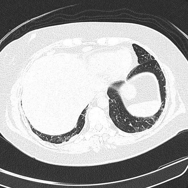
Description: CT scan lungs. Right upper lobe subpleural mass, which measures 3,2 cm, and airspace consolidation at the lateral segment of the middle lobe, associated with several scattered nodules distributed in both lungs, predominantly in the right lung. There are hilar and mediastinal lymphadenopathy, as well as enlarged lymph nodes in the axillary regions and pectoral chains. No pericardial or pleural effusion. Conclusion - Cryptococcus neoformans, with negative results for acid-fast bacilli and malignancy.
Licensing
| This work is licensed under the Creative Commons Attribution-NonCommersial-ShareAlike 4.0 License. |
File history
Click on a date/time to view the file as it appeared at that time.
| Date/Time | Thumbnail | Dimensions | User | Comment | |
|---|---|---|---|---|---|
| current | 06:55, 7 June 2021 |  | 1,778 × 1,778 (882 KB) | Whispyhistory (talk | contribs) | Author: Case courtesy of Dr Antonio Rodrigues de Aguiar Neto, Radiopaedia.org, rID: 78154 Source: https://radiopaedia.org/cases/pulmonary-cryptococcosis-11?lang=gb Description: CT scan lungs. Right upper lobe subpleural mass, which measures 3,2 cm, and airspace consolidation at the lateral segment of the middle lobe, associated with several scattered nodules distributed in both lungs, predominantly in the right lung. There are hilar and mediastinal lymphadenopathy, as well as enlarged lymp... |
You cannot overwrite this file.
File usage
The following file is a duplicate of this file (more details):
- File:Pulmonary cryptococcosis (Radiopaedia 78154-90695 Axial 10).jpg from a shared repository
There are no pages that use this file.