File:Paracoccidioidomycosis-3.jpg

Original file (1,435 × 993 pixels, file size: 353 KB, MIME type: image/jpeg)
Summary
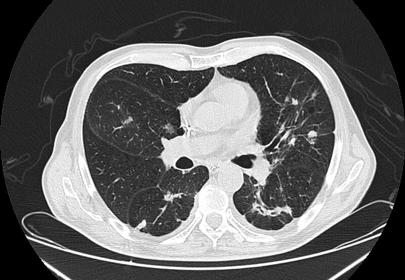
Author: Case courtesy of Dr Thiago Andre Adame, Radiopaedia.org, rID: 65304 Source: https://radiopaedia.org/cases/paracoccidioidomycosis-3?lang=us Description: Ct-scan chest, Multiple irregular nodules, some with internal cavitation, predominantly distributed throughout the posterior regions of upper and middle portions. There is a focal area of ground-glass attenuation with surrounding crescent or ring shaped consolidation (reversed halo sign). Architectural distortion and fibrosis in peripheral and posterior regions of lower lobes. There is also centrolobular emphysema. This case illustrates the lung involvement in a confirmed case of paracoccidioidomycosis, which is a systemic fungal infection endemic in South America.
Licensing
| This work is licensed under the Creative Commons Attribution-NonCommersial-ShareAlike 4.0 License. |
File history
Click on a date/time to view the file as it appeared at that time.
| Date/Time | Thumbnail | Dimensions | User | Comment | |
|---|---|---|---|---|---|
| current | 10:46, 13 July 2021 |  | 1,435 × 993 (353 KB) | Whispyhistory (talk | contribs) | Author: Case courtesy of Dr Thiago Andre Adame, Radiopaedia.org, rID: 65304 Source: https://radiopaedia.org/cases/paracoccidioidomycosis-3?lang=us Description: Ct-scan chest, Multiple irregular nodules, some with internal cavitation, predominantly distributed throughout the posterior regions of upper and middle portions. There is a focal area of ground-glass attenuation with surrounding crescent or ring shaped consolidation (reversed halo sign). Architectural distortion and fibrosis in periph... |
You cannot overwrite this file.
File usage
The following file is a duplicate of this file (more details):
- File:Paracoccidioidomycosis (Radiopaedia 65304-74330 Axial 1).jpg from a shared repository
There are no pages that use this file.