File:PMC5006550 dp0603a07g005a.png
Jump to navigation
Jump to search
No higher resolution available.
PMC5006550_dp0603a07g005a.png (512 × 288 pixels, file size: 291 KB, MIME type: image/png)
File history
Click on a date/time to view the file as it appeared at that time.
| Date/Time | Thumbnail | Dimensions | User | Comment | |
|---|---|---|---|---|---|
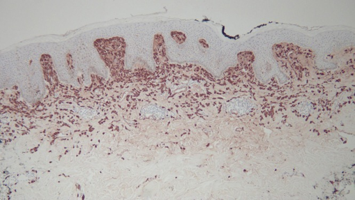
| current | 21:26, 8 March 2022 | | 512 × 288 (291 KB) | Ozzie10aaaa | Author:Philip R. Cohen,Department of Dermatology, University of California San Diego(Openi/National Library of Medicine) Source:https://openi.nlm.nih.gov/detailedresult?img=PMC5006550_dp0603a07g005a&query=Solitary%20mastocytoma&it=xg&req=4&npos=2 Description:f5-dp0603a07: Distant (a) and closer (b and c) views of the right abdomen mastocytoma skin biopsy, stained with tryptase, show that the cells in the upper dermis are highlighted by the immunoperoxidase stain [tryptase, a = x4; b = x20; c=... |
File usage
There are no pages that use this file.