
No higher resolution available.
This file is from a shared repository and may be used by other projects.
The description on its file description page there is shown below.
License
CC0 1.0 Universal (CC0 1.0) Public Domain Dedication
- &
Attribution 4.0 International (CC BY 4.0)
Summary
Author:Thaís Fenz Araujo, Erlane Marques Ribeiro, Anderson Pontes Arruda, Carolina Araujo Moreno, Paula Frassinetti Vasconcelos de Medeiros, Renata Moldenhauer Minillo, Débora Gusmão Melo, Chong Ae Kim, Maria Juliana Rodovalho Doriqui, Têmis Maria Felix, Rodrigo Ambrosio Fock, Denise Pontes Cavalcanti,Skeletal Dysplasia Group, Department of Medical Genetics, Faculty of Medical Sciences, State University of Campinas,Children’s Hospital Albert Sabin, Medical Sciences Faculty of Juazeiro do Norte (FMJ), Perinatal Genetics Program, Department of Medical Genetics, Faculty of Medical Sciences, State University of Campinas, Federal University of Campina Grande, Children’s Clinic, City Hall of Guarulhos, Medical Department, Federal University of de São Carlos (UFSCAR),Medical Genetics Unit, Children’s Institute, Medical Sciences Faculty, University of São Paulo (FCMUSP), Children’s Hospital Juvêncio Mattos, Medical Genetics Service, Clinical Hospital of Porto Alegre, Centro de Genética Médica da Universidade Federal de São Paulo(Openi/National Library of Medicine) Source:https://openi.nlm.nih.gov/detailedresult?img=PMC4997772_40001_2016_228_Fig2_HTML&query=Pycnodysostosis&it=xg&req=4&npos=2
Description:Fig2: Clinical and radiological findings of two patients affected by pycnodysostosis. a–b The typical facial appearance of the pycnodysostosis (large frontal, midfacial hypoplasia, low set ears, and beaked nose), here observed in the patient 27. c–d Cranial X-rays of patient 27 showing increased bone density, opened sutures, parietal bossing and obtuse mandibular angle. e The hands of patient 27 showing brachydactyly. f X-rays of the hands of the patient 2 showing increased bone density, brachydactyly and acroosteolysis of the distal phalanges, mainly observed in both second fingers
File history
Click on a date/time to view the file as it appeared at that time.
| Date/Time | Thumbnail | Dimensions | User | Comment |
|---|
| current | 22:55, 2 January 2022 | 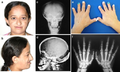 | 512 × 307 (256 KB) | Ozzie10aaaa | Author:Thaís Fenz Araujo, Erlane Marques Ribeiro, Anderson Pontes Arruda, Carolina Araujo Moreno, Paula Frassinetti Vasconcelos de Medeiros, Renata Moldenhauer Minillo, Débora Gusmão Melo, Chong Ae Kim, Maria Juliana Rodovalho Doriqui, Têmis Maria Felix, Rodrigo Ambrosio Fock, Denise Pontes Cavalcanti,Skeletal Dysplasia Group, Department of Medical Genetics, Faculty of Medical Sciences, State University of Campinas,Children’s Hospital Albert Sabin, Medical Sciences Faculty of Juazeiro do Nort... |
File usage
The following page uses this file: