
No higher resolution available.
This file is from a shared repository and may be used by other projects.
The description on its file description page there is shown below.
License
Attribution-NonCommercial-NoDerivatives 4.0 International (CC BY-NC-ND 4.0)
Summary
Author:Musil IL, Gilbertson-Dahdal D, Elliott SP, University of Arizona, College of Medicine, Department of Radiology, University of Arizona Health Sciences Center,Section of Pediatric Infectious Diseases, University of Arizona Health Sciences Center (Openi/National Library of Medicine) Source:https://openi.nlm.nih.gov/detailedresult?img=PMC4897600_fx2a&query=Disseminated%20coccidioidomycosis&it=xg&req=4&npos=3 Description:f20: 16-year-old female with disseminated coccidioidomycosis. CT of the chest and abdomen with contrast. (A) Multiple necrotic portocaval lymph nodes are shown here. Mild narrowing of the proximal superior vena cava by the necrotic mass is seen within the mediastinum. A necrotic enhancing soft-tissue mass occupies the majority of the mediastinum above the heart. This mass represents a confluence of necrotic lymphadenopathy. It is compressing the superior vena cava proximally, but the vessel remains patent. The conglomerate soft-tissue mass in the mediastinum is also pushing the pulmonary artery and aorta to the left, causing elongation and narrowing of the right pulmonary artery, and is partially compressing the right bronchus as well. (B) This chest CT demonstrates numerous hypodense nodules in the liver and in the spleen that represent foci of coccidioidomycosis infection. The abnormal enhancement of the liver is likely related to SVC compression. (C) Multiple cystic structures demonstrated within the spleen.
File history
Click on a date/time to view the file as it appeared at that time.
| Date/Time | Thumbnail | Dimensions | User | Comment |
|---|
| current | 19:09, 14 March 2022 | 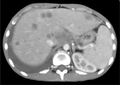 | 512 × 361 (127 KB) | Ozzie10aaaa | Author:Musil IL, Gilbertson-Dahdal D, Elliott SP, University of Arizona, College of Medicine, Department of Radiology, University of Arizona Health Sciences Center,Section of Pediatric Infectious Diseases, University of Arizona Health Sciences Center (Openi/National Library of Medicine) Source:https://openi.nlm.nih.gov/detailedresult?img=PMC4897600_fx2a&query=Disseminated%20coccidioidomycosis&it=xg&req=4&npos=3 Description:f20: 16-year-old female with disseminated coccidioidomycosis. CT of th... |
File usage
There are no pages that use this file.