File:PMC4789832 SCIENTIFICA2016-2173427 (1).png

From WikiProjectMed
Jump to navigation Jump to search

PMC4789832_SCIENTIFICA2016-2173427_(1).png(257 × 144 pixels, file size: 92 KB, MIME type: image/png)

This file is from a shared repository and may be used by other projects. The description on its file description page there is shown below.

Summary

Description
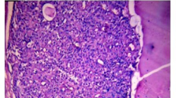
English: fig1: (a) Nodular hidradenoma, showing clear and polygonal cells with foci of eosinophilic material (H&E, ×100). (b) Hidradenoma papilliferum, showing scattered cystic and branching spaces with numerous papillary projections lined by double layer of epithelium (H&E, ×100). (c) Eccrine spiradenoma, showing well circumscribed aggregates of tumour cells in dermis (H&E, ×100). (d) Syringoma, showing numerous small ducts embedded in a fibrous stroma (H&E, ×100).
Date
Source https://openi.nlm.nih.gov/detailedresult?img=PMC4789832_SCIENTIFICA2016-2173427.001&query=Hidradenoma&it=xg&req=4&npos=2
Author Shukla P, Fatima U, Malaviya AK

Licensing

English: This file is licensed CC BY-NC 4.0

This file was uploaded with UploadWizard.

File history

Click on a date/time to view the file as it appeared at that time.

Date/TimeThumbnailDimensionsUserComment
current03:12, 14 January 2023Thumbnail for version as of 03:12, 14 January 2023257 × 144 (92 KB)Ozzie10aaaaUploaded a work by Shukla P, Fatima U, Malaviya AK from https://openi.nlm.nih.gov/detailedresult?img=PMC4789832_SCIENTIFICA2016-2173427.001&query=Hidradenoma&it=xg&req=4&npos=2 with UploadWizard

There are no pages that use this file.