File:PMC4466302 ad-27-353-g002.png
Jump to navigation
Jump to search
No higher resolution available.
PMC4466302_ad-27-353-g002.png (512 × 168 pixels, file size: 164 KB, MIME type: image/png)
File history
Click on a date/time to view the file as it appeared at that time.
| Date/Time | Thumbnail | Dimensions | User | Comment | |
|---|---|---|---|---|---|
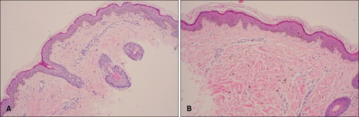
| current | 19:43, 8 March 2022 | 512 × 168 (164 KB) | Ozzie10aaaa | Author:Jun HJ, Kim SM, Cho SH, Lee JD, Kim HS ,Department of Dermatology, Incheon St. Mary's Hospital, College of Medicine, The Catholic University of Korea(Openi/National Library of Medicine) Source:https://openi.nlm.nih.gov/detailedresult?img=PMC4466302_ad-27-353-g002&query=Phakomatosis%20pigmentovascularis&it=xg&req=4&npos=48 Description:F2: (A) Increased melanin pigment in the basal layer and dilation of capillary in the dermis (H&E, ×100). (B) Increased melanin pigment in the basal layer... |
File usage
The following page uses this file: