File:PMC4312899 pim0036-0170-f2.png
Jump to navigation
Jump to search
No higher resolution available.
PMC4312899_pim0036-0170-f2.png (512 × 185 pixels, file size: 225 KB, MIME type: image/png)
Summary
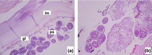
| Description |
English: fig02: Surgical pathology of the cystic echinococcosis. Microscopic examination of the pathological specimen demonstrating the multilayer laminated membrane of the hydatid cyst wall (H&E 200 × ). Section on the left showed a cystic echinococcosis with higher IgG level. The germinal layer (gl) and protoscolices (ps).were enclosed complete with laminated membrane (lm); the section on the right showed a cystic echinococcosis with higher IgG level. The hydatid cyst had ruptured. |
| Date | |
| Source | https://openi.nlm.nih.gov/detailedresult?img=PMC4312899_pim0036-0170-f2&query=Echinococcosis&it=xg&req=4&npos=9 |
| Author | Chen X, Zhang J, Feng X, Chen X, Yin S, Wen H, Zheng S |
Licensing
| This work is licensed under the Creative Commons Attribution 3.0 unported License.
Anyone may use this image for any purpose provided it is attributed as specified. |
 |
This file was uploaded with UploadWizard.
File history
Click on a date/time to view the file as it appeared at that time.
| Date/Time | Thumbnail | Dimensions | User | Comment | |
|---|---|---|---|---|---|
| current | 00:11, 19 November 2022 | 512 × 185 (225 KB) | Ozzie10aaaa | Uploaded a work by Chen X, Zhang J, Feng X, Chen X, Yin S, Wen H, Zheng S from https://openi.nlm.nih.gov/detailedresult?img=PMC4312899_pim0036-0170-f2&query=Echinococcosis&it=xg&req=4&npos=9 with UploadWizard |
File usage
The following page uses this file: