
No higher resolution available.
This file is from a shared repository and may be used by other projects.
The description on its file description page there is shown below.
License
Attribution 3.0 Unported (CC BY 3.0)
Summary
Author:Brunetti-Pierri N, Torrado M, Fernandez Mdel C, Tello AM, Arberas CL, Cardinale A, Piccolo P, Bacino CA,Telethon Institute of Genetics and Medicine Naples, Italy ; Department of Translational Medicine, Federico II University of Naples Naples, Italy (Openi/National Library of Medicine) Source:https://openi.nlm.nih.gov/detailedresult?img=PMC4303216_mgg30002-0467-f1&query=Terminal%20osseous%20dysplasia%20with%20pigmentary%20defects&it=xg&req=4&npos=1 Description: fig01: (A) Pedigree 1 family. Patients II.2 and II.4 are depicted. Note facial hyperpigmented spots, atrophic lesions of the temporal region, oral frenulae, brachydactyly and camptodactyly, and digital fibromas (arrows). Feet of patient II.2 showed variable shortening of second through V toes, with elongated first toes bilaterally. Hand X-rays of patient II.2 showed in the left hand a short and wide III metacarpal, thin IV metacarpal, short V metacarpal, mild hypoplasia of all middle phalanges, hypoplasia of all distal phalanges, fusion of the III carpal-metacarpal, joint angulation; in the right hand thin II metacarpal, slight hypoplastic tufts. The thumbs are spared. The face of patient II.4 shows atrophic lesions with hyperpigmentation (arrow). Hands of patient II.4 have periungueal fibromatosis lesions over the fourth digit (arrows). Hand X-rays of patient II.4 show delayed carpal center ossification; the III metacarpal is hypoplastic and widened (bullet-shaped); angulated middle phalanges-distal phalanges joint of the III digit is noted. Foot X-rays of patient II.4 show asymmetrical involvement; on the right foot the II and IV metatarsals are hypoplastic or have an amorphous shape, the great toes are elongated. On the far right, a picture of both girls' mother is shown (I.2) with multiple frenulae in the lower lip (arrow). (B) Pedigree of family 2 is shown. Frontal and profile views of the face of patient II.2 with hyperpigmented atrophic lesions over malar and temporal regions (arrows). Right arm showed mild pterygia and brachydactyly on both hands mostly affecting first and third through V metacarpals. Camptodactyly is also present. On the lower figure, individual I.2 is shown. Note her multiple frenulae (arrow) and brachydactyly. (C) Sanger sequencing shows the c.5217G>A mutation in the index case (II.2 from pedigree 1 [A]) and normal sequence in her mother (I.2).
File history
Click on a date/time to view the file as it appeared at that time.
| Date/Time | Thumbnail | Dimensions | User | Comment |
|---|
| current | 15:44, 5 August 2021 | 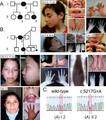 | 512 × 581 (564 KB) | Ozzie10aaaa | Author:Brunetti-Pierri N, Torrado M, Fernandez Mdel C, Tello AM, Arberas CL, Cardinale A, Piccolo P, Bacino CA,Telethon Institute of Genetics and Medicine Naples, Italy ; Department of Translational Medicine, Federico II University of Naples Naples, Italy (Openi/National Library of Medicine) Source:https://openi.nlm.nih.gov/detailedresult?img=PMC4303216_mgg30002-0467-f1&query=Terminal%20osseous%20dysplasia%20with%20pigmentary%20defects&it=xg&req=4&npos=1 Description: fig01: (A) Pedigree 1 fa... |
File usage
There are no pages that use this file.