File:PMC4000873 kjtcs-47-145-g003.png
Jump to navigation
Jump to search
No higher resolution available.
PMC4000873_kjtcs-47-145-g003.png (512 × 191 pixels, file size: 184 KB, MIME type: image/png)
File history
Click on a date/time to view the file as it appeared at that time.
| Date/Time | Thumbnail | Dimensions | User | Comment | |
|---|---|---|---|---|---|
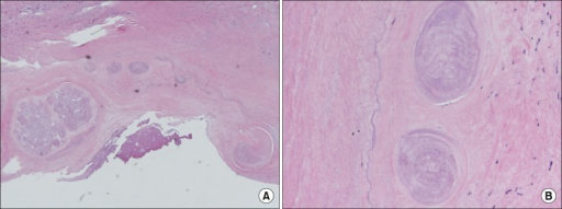
| current | 20:59, 2 April 2022 | 512 × 191 (184 KB) | Ozzie10aaaa | Author:Bang SH, Park JB, Chee HK, Kim JS, Ko SM, Kim WS, Shin JK, Department of Thoracic and Cardiovascular Surgery, Konkuk University School of Medicine(Openi/National Library of Medicine) Source:https://openi.nlm.nih.gov/detailedresult?img=PMC4000873_kjtcs-47-145-g003&query=Trichinosis&it=xg&req=4&npos=16 Description:F3: The larvae of Trichinella spiralis are seen in the fibromuscular tissue of the right ventricle. (A) H&E, ×40. (B) H&E, ×200. |
File usage
The following page uses this file: