File:PMC3820176 jpts-25-1209-g001.png
Jump to navigation
Jump to search
No higher resolution available.
PMC3820176_jpts-25-1209-g001.png (512 × 275 pixels, file size: 136 KB, MIME type: image/png)
File history
Click on a date/time to view the file as it appeared at that time.
| Date/Time | Thumbnail | Dimensions | User | Comment | |
|---|---|---|---|---|---|
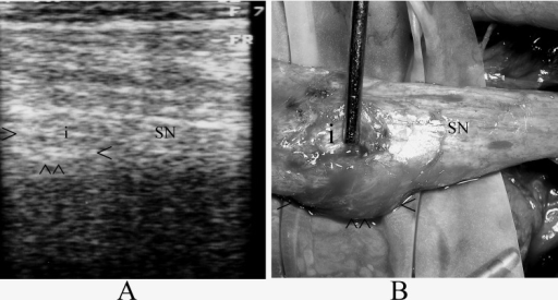
| current | 19:45, 4 November 2021 | | 512 × 275 (136 KB) | Ozzie10aaaa | Author:Bilgici A, Cokluk C, Aydın K, Department of Physical Therapy and Rehabilitation, Ondokuzmayıs University (Openi/National Library of Medicine) Source: https://openi.nlm.nih.gov/detailedresult?img=PMC3820176_jpts-25-1209-g001&query=sciatic%20nerve%20injury&it=xg&req=4&npos=88 Description:fig_001: A. Sonographic examination revealed the injury site, neuroma formation and sciaticnerve (arrowheads show neuroma formation; i: injury site; SN: sciatic nerve). B.Intraoperative photograph shows... |
File usage
The following page uses this file: