File:PMC3693162 JCE2013-549041.002.png
Jump to navigation
Jump to search

Size of this preview: 191 × 600 pixels. Other resolutions: 76 × 240 pixels | 326 × 1,024 pixels.
Original file (326 × 1,024 pixels, file size: 772 KB, MIME type: image/png)
File history
Click on a date/time to view the file as it appeared at that time.
| Date/Time | Thumbnail | Dimensions | User | Comment | |
|---|---|---|---|---|---|
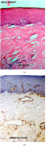
| current | 20:43, 18 April 2022 | 326 × 1,024 (772 KB) | Ozzie10aaaa | Author:Nayak MT, Singh A, Desai RS, Vanaki SS, Department of Oral & Maxillofacial Pathology, Vyas Dental College and Hospital(Openi/National Library of Medicine) Source:https://openi.nlm.nih.gov/detailedresult?img=PMC3693162_JCE2013-549041.002&query=Oral%20submucous%20fibrosis&it=xg&req=4&npos=1 Description:fig2: (a) Mild cases of Oral submucous fibrosis under H/E staining. (b) Mild cases of oral submucous fibrosis under vimentin immunohistochemical staining. Note the staining intensity of vi... |
File usage
There are no pages that use this file.