
No higher resolution available.
This file is from a shared repository and may be used by other projects.
The description on its file description page there is shown below.
License
Attribution 2.0 Generic (CC BY 2.0)
Summary
Author:Zaidan M, Mariotte E, Galicier L, Arnulf B, Meignin V, Vérine J, Mahr A, Azoulay E ,Department of Medical Intensive Care Unit, Hôpital Saint-Louis (Openi/National Library of Medicine) Source:https://openi.nlm.nih.gov/detailedresult?img=PMC3488028_2110-5820-2-31-1&query=Cryoglobulinemic%20vasculitis&it=xg&req=4&npos=1 Description:F1: Widespread fulminant cryoglobulinemic vasculitis is defined as involvement of the skin and at least two other organs, such as illustrated by the following case. A 50-year-old woman presented with fever, bilateral purpuric lesions, dyspnea, and acute kidney injury. The bronchoalveolar lavage showed severe hemorrhage with predominance of macrophages and neutrophils (a, May-Grünwald stain, x200). The skin biopsy revealed typical features of leukocytoclastic vasculitis with intraluminal thrombosis and fibrinoid-type necrosis associated with infiltration by neutrophils of the vessel walls in the dermis (b, hematoxylin-eosin-saffron stain, x200). The kidney biopsy displayed membranoproliferative glomerulonephritis. The glomeruli showed mesangial expansion by increased mesangial cell number and matrix, and some intracapillary "protein thrombi" (c, Masson’s trichrome stain, x400). A mild endocapillary hypercellularity and duplication of the glomerular basement membrane with “double contours” were also observed (d, Jones stain, x400). Immunofluorescence study showed granular IgG-positive deposits with lambda light-chain isotype restriction localized to the glomerular capillary wall, mesangium, and intracapillary thrombi (e, IgG Immunofluorescence staining, x400, lambda staining not shown). IgM, IgA, and kappa staining were all negative (data not shown). Lambda staining was comparable to IgG staining (data not shown), which was consistent with renal involvement of type I cryoglobulinemia (IgG lambda). Finally, the diagnosis was widespread cryoglobulinemic vasculitis with skin, lung, and kidney involvement secondary to monoclonal gammopathy of undetermined significance associated with Sjögren’s syndrome.
File history
Click on a date/time to view the file as it appeared at that time.
| Date/Time | Thumbnail | Dimensions | User | Comment |
|---|
| current | 18:12, 12 October 2021 | 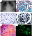 | 512 × 580 (623 KB) | Ozzie10aaaa | Author:Zaidan M, Mariotte E, Galicier L, Arnulf B, Meignin V, Vérine J, Mahr A, Azoulay E ,Department of Medical Intensive Care Unit, Hôpital Saint-Louis (Openi/National Library of Medicine) Source:https://openi.nlm.nih.gov/detailedresult?img=PMC3488028_2110-5820-2-31-1&query=Cryoglobulinemic%20vasculitis&it=xg&req=4&npos=1 Description:F1: Widespread fulminant cryoglobulinemic vasculitis is defined as involvement of the skin and at least two other organs, such as illustrated by the following... |
File usage
The following page uses this file: