File:PMC3271412 NAJMS-3-527-g002.png
Jump to navigation
Jump to search

Size of this preview: 451 × 599 pixels. Other resolutions: 181 × 240 pixels | 512 × 680 pixels.
Original file (512 × 680 pixels, file size: 724 KB, MIME type: image/png)
File history
Click on a date/time to view the file as it appeared at that time.
| Date/Time | Thumbnail | Dimensions | User | Comment | |
|---|---|---|---|---|---|
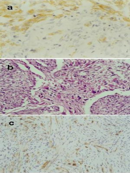
| current | 22:36, 5 January 2022 | | 512 × 680 (724 KB) | Ozzie10aaaa | Author:Andaloussi-Saghir K, Oukabli M, El Marjany M, Sifat H, Hadadi K, Mansouri H ,Department of Radiotherapy Oncology, Mohammed V Military Hospital(Openi/National Library of medicine)Source:https://openi.nlm.nih.gov/detailedresult?img=PMC3271412_NAJMS-3-527-g002&query=&req=4 Description:F2: Photomicrographs of the tumor. a: The gliomatous component GFAP positive (×100). b: Tumour characterised by a biphasic tissue pattern with alternating areas displaying glial and mesenchymal differentiati... |
File usage
The following page uses this file: