File:PMC2699843 TOOPHTJ-2-165 F1 (1) (1).png

From WikiProjectMed
Jump to navigation Jump to search

PMC2699843_TOOPHTJ-2-165_F1_(1)_(1).png(214 × 171 pixels, file size: 17 KB, MIME type: image/png)

This file is from a shared repository and may be used by other projects. The description on its file description page there is shown below.

Summary

Description
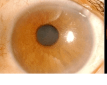
English: F1: (a) Slit-lamp examination showed corneal edema and rubeosis iridis in the left eye. (b) Fundus examination showed glaucomatous changes of the optic disc and almost normal retinal vessels. (c) Three-dimensional computed tomography angiography revealed absence of the left internal carotid artery and thinned common carotid artery (arrows). (d) A few days after the trabeculectomy, rubeosis iridis disappeared.
Date
Source https://openi.nlm.nih.gov/detailedresult?img=PMC2699843_TOOPHTJ-2-165_F1&query=Rubeosis%20iridis&it=xg&req=4&npos=3
Author Gyotoku Y, Kawaji T, Inatani M, Fukushima M, Tanihara H

Licensing

English: This file is licensed CC BY-NC 3.0

This file was uploaded with UploadWizard.

File history

Click on a date/time to view the file as it appeared at that time.

Date/TimeThumbnailDimensionsUserComment
current02:34, 24 August 2022Thumbnail for version as of 02:34, 24 August 2022214 × 171 (17 KB)Ozzie10aaaaUploaded a work by Gyotoku Y, Kawaji T, Inatani M, Fukushima M, Tanihara H from https://openi.nlm.nih.gov/detailedresult?img=PMC2699843_TOOPHTJ-2-165_F1&query=Rubeosis%20iridis&it=xg&req=4&npos=3 with UploadWizard

There are no pages that use this file.