File:PMC2648628 IPID2008-750479.007 (1).png

From WikiProjectMed
Jump to navigation Jump to search

PMC2648628_IPID2008-750479.007_(1).png(484 × 201 pixels, file size: 157 KB, MIME type: image/png)

This file is from a shared repository and may be used by other projects. The description on its file description page there is shown below.

Summary

Description
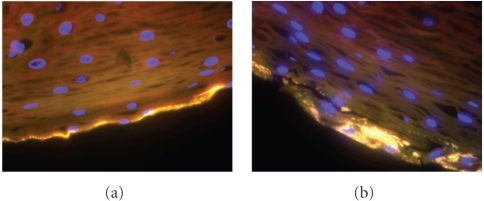
English: fig7: Vaginal biopsy from asubject with BV. A Gardnerella vaginalis biofilm (yellow) is detected at the edge ofthe vaginal epithelium (bottom) by fluorescence in situ hybridization(FISH). The yellow color is the resultof using a combination of probes targeting G. vaginalis (Red), all bacteria (Eub338, green), and 4′,6-diamidine-2-phenylindole, dihydrochloride(DAPI, blue) which stains DNA. Notehuman cell nuclei in blue. The image onthe right shows a vaginal epithelial cell with a cluster of G. vaginalis breaking off the epitheliumand likely forming a clue cell.
Date
Source https://openi.nlm.nih.gov/detailedresult?img=PMC2648628_IPID2008-750479.007&query=Gardnerella%20vaginalis&it=xg&req=4&npos=6
Author Srinivasan S, Fredricks DN

Licensing

English: This file is licensed CC BY-NC 4.0

This file was uploaded with UploadWizard.

File history

Click on a date/time to view the file as it appeared at that time.

Date/TimeThumbnailDimensionsUserComment
current22:09, 28 September 2023Thumbnail for version as of 22:09, 28 September 2023484 × 201 (157 KB)Ozzie10aaaaUploaded a work by Srinivasan S, Fredricks DN from https://openi.nlm.nih.gov/detailedresult?img=PMC2648628_IPID2008-750479.007&query=Gardnerella%20vaginalis&it=xg&req=4&npos=6 with UploadWizard

There are no pages that use this file.