File:PMC2627172 kjr-9-91-g001 (1).png
Jump to navigation
Jump to search
No higher resolution available.
PMC2627172_kjr-9-91-g001_(1).png (497 × 548 pixels, file size: 122 KB, MIME type: image/png)
.png)
File history
Click on a date/time to view the file as it appeared at that time.
| Date/Time | Thumbnail | Dimensions | User | Comment | |
|---|---|---|---|---|---|
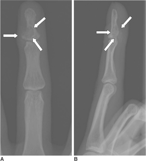
| current | 19:40, 21 April 2022 | | 497 × 548 (122 KB) | Ozzie10aaaa | Author:Choi SJ, Ahn JH, Kang G, Lee JH, Park MS, Ryu DS, Jung SM ,Department of Radiology, Asan Foundation, GangNeung Asan Hospital, University of Ulsan College of Medicine(Openi/National Library of Medicine)Source:https://openi.nlm.nih.gov/detailedresult?img=PMC2627172_kjr-9-91-g001&query=Aponeurotic%20fibroma&it=xg&req=4&npos=3 Description:F1: Left middle finger AP (A) and lateral (B) views show eccentrically located well-defined osteolytic lesion in the base of the distal phalanx (arrows).... |
File usage
There are no pages that use this file.
.png)