File:PMC2526518 jkms-23-551-g003.png
Jump to navigation
Jump to search
No higher resolution available.
PMC2526518_jkms-23-551-g003.png (433 × 263 pixels, file size: 105 KB, MIME type: image/png)
File history
Click on a date/time to view the file as it appeared at that time.
| Date/Time | Thumbnail | Dimensions | User | Comment | |
|---|---|---|---|---|---|
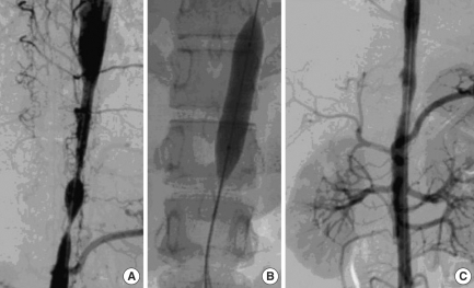
| current | 23:37, 8 November 2021 | | 433 × 263 (105 KB) | Ozzie10aaaa | Author:Shim BJ, Youn HJ, Kim YC, Kim WT, Choi YS, Lee DH, Park C, Oh YS, Chung WS, Kim JH, Choi KB, Hong SJ, Jung SE, Hahn ST,Division of Cardiology, Department of Internal Medicine, College of Medicine, The Catholic University of Korea (Openi/National Library of Medicine) Source:https://openi.nlm.nih.gov/detailedresult?img=PMC2526518_jkms-23-551-g003&query=&req=4 Description:F3: (A) 3D-CT angiography shows focal stenosis in the descending aorta, but not the renal artery. (B) Percutaneous tra... |
File usage
The following page uses this file: