No higher resolution available.
This file is from a shared repository and may be used by other projects.
The description on its file description page there is shown below.
License
Attribution 2.0 Generic (CC BY 2.0)
Summary
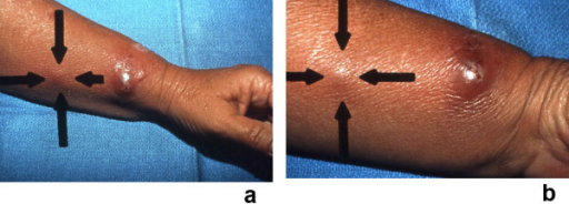
Author:Cohen PR, University of Houston Health Center(Openi/National Library of Medicine) Source:https://openi.nlm.nih.gov/detailedresult?img=PMC1963326_1750-1172-2-34-10&query=Reactive%20neutrophilic%20dermatoses&it=xg&req=4&npos=1 Description:F10: (a and b). Sweet's syndrome with lesions distributed in a sporotrichoid pattern. A 53-year-old Hispanic woman presented to the emergency room with a recent episode of a "sore throat," fever, and a 2-day history of a swollen, tender left wrist accompanied by erythema that was beginning to extend proximally; a bacterial cellulitis was clinically suspected and she was started on oral antibiotics: double-strength trimethoprim-sulfamethoxazole twice daily and 400 mg of ofloxacin twice daily. She was initially seen in the dermatology clinic 9 days later; she was still febrile and her original lesion had developed into a painful larger pseudovesicular nodule on the radial side of her left wrist. In addition, distal (a) and closer (b) views show a smaller red dermal nodule (between arrows) that appeared on her left arm proximal to the original lesion. The clinical differential diagnosis included infections whose lesions demonstrated a sporotrichoid pattern (sporotrichosis and atypical mycobacterial infection) and Sweet's syndrome. Biopsies for microscopic and culture evaluation were performed. In addition to her antibiotics, the patient was started on oral saturated solution of potassium iodide (3 drops 3 times each day and increased by 1 drop each day to a final dose of 10 drops 3 times each day). The hematoxylin and eosin-stained sections from her biopsy showed a neutrophilic dermatosis; the bacterial, mycobacterial, and fungal cultures were negative for organisms. Within a few days after initiating treatment with potassium iodide, her symptoms resolved and her skin lesions began to improve. (From [23] Cohen PR, Kurzrock R: Sweet's syndrome: a neutrophilic dermatosis classically associated with acute onset and fever. Clin Dermatol 2000;18:265–282. Copyright 2000, Reprinted with permission from Elsevier Ltd, Oxford, United Kingdom.)
File history
Click on a date/time to view the file as it appeared at that time.
| Date/Time | Thumbnail | Dimensions | User | Comment |
|---|
| current | 20:46, 31 March 2022 |  | 512 × 184 (225 KB) | Ozzie10aaaa | Author:Cohen PR, University of Houston Health Center(Openi/National Library of Medicine) Source:https://openi.nlm.nih.gov/detailedresult?img=PMC1963326_1750-1172-2-34-10&query=Reactive%20neutrophilic%20dermatoses&it=xg&req=4&npos=1 Description:F10: (a and b). Sweet's syndrome with lesions distributed in a sporotrichoid pattern. A 53-year-old Hispanic woman presented to the emergency room with a recent episode of a "sore throat," fever, and a 2-day history of a swollen, tender left wrist accom... |
File usage
There are no pages that use this file.