File:Laugier-Hunziker Syndrome histology.png
Jump to navigation
Jump to search
Laugier-Hunziker_Syndrome_histology.png (512 × 455 pixels, file size: 625 KB, MIME type: image/png)
Summary
Author:Barman PD, Das A, Mondal AK, Kumar P.
Source:"Laugier-Hunziker Syndrome Revisited". Indian J Dermatol. 2016 May-Jun;61(3):338-9. doi: 10.4103/0019-5154.182429. PMID: 27293265; PMCID: PMC4885197. Via Openi
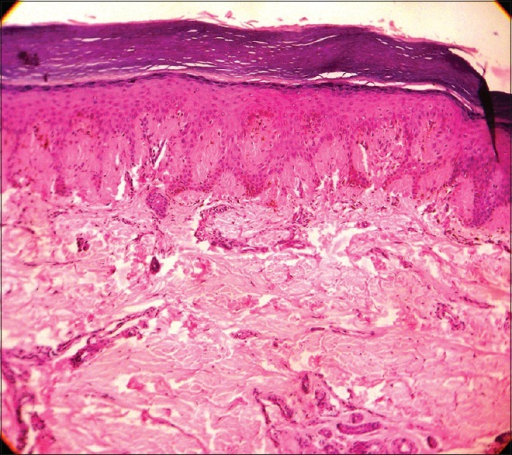
Description: Laugier-Hunziker Syndrome melanonychia. Photomicrograph showing hyperkeratosis, mild acanthosis, interwoven rete ridges, increased basal layer pigmentation with an increased number of normal melanocytes (H and E, ×10)
Licensing
File history
Click on a date/time to view the file as it appeared at that time.
| Date/Time | Thumbnail | Dimensions | User | Comment | |
|---|---|---|---|---|---|
| current | 10:53, 13 April 2022 |  | 512 × 455 (625 KB) | Whispyhistory (talk | contribs) | == Summary == '''Author''':Barman PD, Das A, Mondal AK, Kumar P. '''Source''':[https://www.ncbi.nlm.nih.gov/pmc/articles/PMC4885197/ "Laugier-Hunziker Syndrome Revisited"]. Indian J Dermatol. 2016 May-Jun;61(3):338-9. doi: 10.4103/0019-5154.182429. PMID: 27293265; PMCID: PMC4885197. [https://openi.nlm.nih.gov/detailedresult?img=PMC4885197_IJD-61-338-g001&query=melanonychia&it=xg&req=4&npos=23 Via Openi] '''Description''': Laugier-Hunziker Syndrome melanonychia. Photomicrograph showing hype... |
You cannot overwrite this file.
File usage
There are no pages that use this file.
