File:Influenza virus particle 8430 lores.jpg
Original file (1,663 × 1,423 pixels, file size: 763 KB, MIME type: image/jpeg)
Summary
| Description |
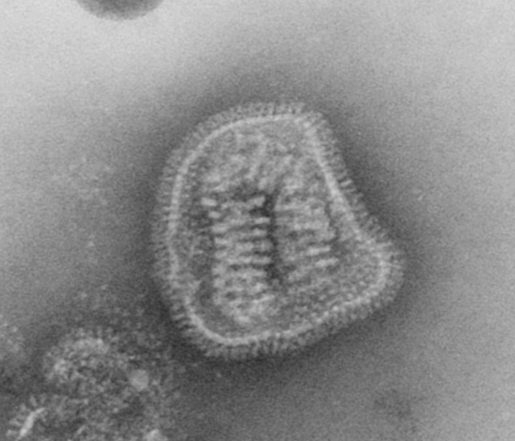
English: This negative-stained transmission electron micrograph (TEM) depicts the ultrastructural details of an influenza virus particle, or “virion”. A member of the taxonomic family Orthomyxoviridae, the influenza virus is a single-stranded RNA organism
The flu is a contagious respiratory illness caused by influenza viruses. It can cause mild to severe illness, and at times can lead to death. The best way to prevent this illness is by getting a flu vaccination each fall. Every year in the United States, on average: - 5% to 20% of the population gets the flu - more than 200,000 people are hospitalized from flu complications, and - about 36,000 people die from flu. Some people, such as older people, young children, and people with certain health conditions, are at high risk for serious flu complications. See PHIL 10073 for a colorized version of this image. Influenza A and B are the two types of influenza viruses that cause epidemic human disease. Influenza A viruses are further categorized into subtypes on the basis of two surface antigens: hemagglutinin and neuraminidase. Influenza B viruses are not categorized into subtypes. Since 1977, influenza A (H1N1) viruses, influenza A (H3N2) viruses, and influenza B viruses have been in global circulation. In 2001, influenza A (H1N2) viruses that probably emerged after genetic reassortment between human A (H3N2) and A (H1N1) viruses began circulating widely. Both influenza A and B viruses are further separated into groups on the basis of antigenic characteristics. New influenza virus variants result from frequent antigenic change (i.e., antigenic drift) resulting from point mutations that occur during viral replication. Influenza B viruses undergo antigenic drift less rapidly than influenza A viruses.Deutsch: Ein behülltes Virus aus der Gattung Influenzavirus in einer TEM-Aufnahme: Acht helikale Kapside werden von einer Virushülle umschlossen (Partikel ca. 80−120 nm im Durchmesser).
Français : Virus de la grippe en microscopie électronique.
Čeština: Virus chřipky pod elektronovým mikroskopem - na povrchu je patrná neuraminidáza a hemaglutinin.
Српски / srpski: Вирус грипа - велико увећање. |
||
| Date | |||
| Source |
|
||
| Author |
|
||
| Permission (Reusing this file) |
PD-USGov-HHS-CDC English: None - This image is in the public domain and thus free of any copyright restrictions. As a matter of courtesy we request that the content provider be credited and notified in any public or private usage of this image. |
||
| Other versions |
|

Licensing
This image is a work of the Centers for Disease Control and Prevention, part of the United States Department of Health and Human Services, taken or made as part of an employee's official duties. As a work of the U.S. federal government, the image is in the public domain.
|
File history
Click on a date/time to view the file as it appeared at that time.
| Date/Time | Thumbnail | Dimensions | User | Comment | |
|---|---|---|---|---|---|
| current | 08:45, 19 November 2010 | | 1,663 × 1,423 (763 KB) | commons>Masur | original resolution |
File usage
There are no pages that use this file.