File:Giant-cell-tumour-distal-radius-3.PNG

Original file (554 × 944 pixels, file size: 474 KB, MIME type: image/png)
Summary
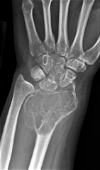
Author: Case courtesy of Dr Matt A. Morgan, Radiopaedia.org, rID: 811
Source: https://radiopaedia.org/cases/giant-cell-tumour-distal-radius?lang=us
Description: The expansile lytic lesion has increased in size and now extends through the entire radius. The lesion has the characteristic appearance of a giant cell tumor. On resection this was found to be a giant cell tumor of bone, extending to subchondral region and eroding through the cortex into surrounding soft tissues.
Licensing
| This work is licensed under the Creative Commons Attribution-NonCommersial-ShareAlike 4.0 License. |
File history
Click on a date/time to view the file as it appeared at that time.
| Date/Time | Thumbnail | Dimensions | User | Comment | |
|---|---|---|---|---|---|
| current | 05:23, 10 May 2021 | | 554 × 944 (474 KB) | Whispyhistory (talk | contribs) | Author: Case courtesy of Dr Matt A. Morgan, Radiopaedia.org, rID: 811 Source: https://radiopaedia.org/cases/giant-cell-tumour-distal-radius?lang=us Description: The expansile lytic lesion has increased in size and now extends through the entire radius. The lesion has the characteristic appearance of a giant cell tumor. On resection this was found to be a giant cell tumor of bone, extending to subchondral region and eroding through the cortex into surrounding soft tissues. |
You cannot overwrite this file.
File usage
The following file is a duplicate of this file (more details):
- File:Giant cell tumor - distal radius (Radiopaedia 81198-94842 Frontal 1).png from a shared repository
.png)
There are no pages that use this file.