File:Epidermis-delimited.JPG
Jump to navigation
Jump to search

Size of this preview: 800 × 496 pixels. Other resolutions: 320 × 198 pixels | 640 × 397 pixels | 1,000 × 620 pixels.
Original file (1,000 × 620 pixels, file size: 240 KB, MIME type: image/jpeg)
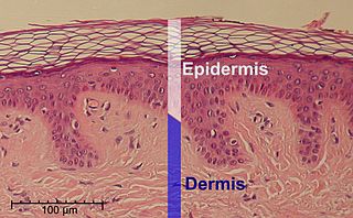
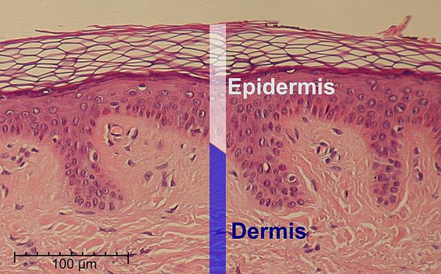
| Description | This is a hematoxylin and eosin stained slide at 10x of normal epidermis. | ||
| Date | |||
| Source |
Scale at lower left was created from the an estimation of mean epidermal cell nuclei of 8.6 μm according to the following study:
|
||
| Author | Cropped and labeled by Fama Clamosa (talk) and Mikael Häggström, respectively | ||
| Permission (Reusing this file) |
I, the copyright holder of this work, hereby publish it under the following license:
|
| This is a retouched picture, which means that it has been digitally altered from its original version. Modifications: cropped. The original can be viewed here: Normal Epidermis and Dermis with Intradermal Nevus 10x.JPG:
|
File history
Click on a date/time to view the file as it appeared at that time.
| Date/Time | Thumbnail | Dimensions | User | Comment | |
|---|---|---|---|---|---|
| current | 16:48, 31 May 2014 | | 1,000 × 620 (240 KB) | commons>Mikael Häggström | Corrected scale |
File usage
There are no pages that use this file.