File:Cryptococcal-ventriculitis.jpg
Cryptococcal-ventriculitis.jpg (558 × 551 pixels, file size: 60 KB, MIME type: image/jpeg)
Summary
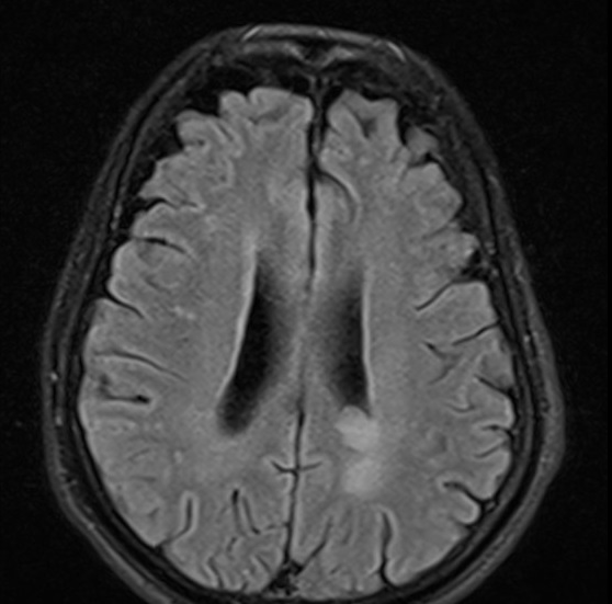
Author: Case courtesy of Dr Sathiyaseelan Maniharan, Radiopaedia.org, rID: 52316 Source: https://radiopaedia.org/cases/cryptococcal-ventriculitis?lang=gb Description: MRI Brain showing Cryptococcal ventriculitis. 55 year old male:A known diabetic patient presented with sudden onset of fever and headache. There is subependymal periventricular hyperintensity along the walls of the lateral and third ventricles on the T2 (not shown) and FLAIR sequences. The ventricular walls show diffusion restriction. The choroid plexus and ventricular walls show contrast enhancement. There is no basal meningeal contrast enhancement. The rest of the brain MRI is normal. The features are consistent with ventriculitis. Radiopaedia case ID: 52316
Licensing
| This work is licensed under the Creative Commons Attribution-NonCommersial-ShareAlike 4.0 License. |
File history
Click on a date/time to view the file as it appeared at that time.
| Date/Time | Thumbnail | Dimensions | User | Comment | |
|---|---|---|---|---|---|
| current | 15:18, 6 June 2021 |  | 558 × 551 (60 KB) | Whispyhistory (talk | contribs) | Author: Case courtesy of Dr Sathiyaseelan Maniharan, Radiopaedia.org, rID: 52316 Source: https://radiopaedia.org/cases/cryptococcal-ventriculitis?lang=gb Description: MRI Brain showing Cryptococcal ventriculitis. 55 year old male:A known diabetic patient presented with sudden onset of fever and headache. There is subependymal periventricular hyperintensity along the walls of the lateral and third ventricles on the T2 (not shown) and FLAIR sequences. The ventricular walls show diffusion restri... |
You cannot overwrite this file.
File usage
The following file is a duplicate of this file (more details):
- File:Cryptococcal ventriculitis (Radiopaedia 52316-58206 Axial 10).jpg from a shared repository
There are no pages that use this file.
