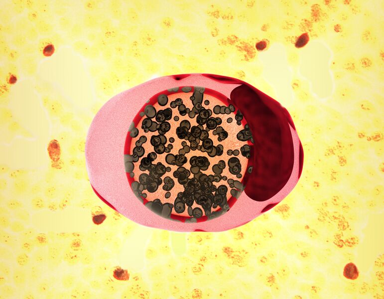
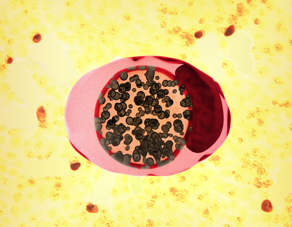
File:53662889580 26f96e4763 o.jpg
Jump to navigation
Jump to search

Size of this preview: 771 × 600 pixels. Other resolutions: 309 × 240 pixels | 617 × 480 pixels | 988 × 768 pixels | 1,280 × 995 pixels | 2,560 × 1,991 pixels | 4,123 × 3,206 pixels.
Original file (4,123 × 3,206 pixels, file size: 4.77 MB, MIME type: image/jpeg)
File history
Click on a date/time to view the file as it appeared at that time.
| Date/Time | Thumbnail | Dimensions | User | Comment | |
|---|---|---|---|---|---|
| current | 22:29, 25 April 2024 | | 4,123 × 3,206 (4.77 MB) | Ozzie10aaaa | Uploaded a work by NIAID and CDC from https://www.flickr.com/photos/niaid/53662889580/ with UploadWizard |
File usage
The following file is a duplicate of this file (more details):
- File:Chlamydia.jpg from a shared repository
The following page uses this file: