File:1-s2nihnihnih.jpg
Jump to navigation
Jump to search
No higher resolution available.
1-s2nihnihnih.jpg (170 × 333 pixels, file size: 18 KB, MIME type: image/jpeg)
File history
Click on a date/time to view the file as it appeared at that time.
| Date/Time | Thumbnail | Dimensions | User | Comment | |
|---|---|---|---|---|---|
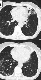
| current | 14:59, 7 October 2023 | | 170 × 333 (18 KB) | Ozzie10aaaa | Uploaded a work by Yuka Aida, Takumi Kiwamoto, Kazutaka Fujita, Hiroaki Ishikawa, Haruna Kitazawa, Hiroko Watanabe, Nobuyuki Hizawa from https://www.sciencedirect.com/science/article/pii/S2213007118302351 with UploadWizard |
File usage
The following page uses this file: