File:1-s2.0-S1930043317306222-radcr476-fig-0002.jpg

From WikiProjectMed
Jump to navigation Jump to search

1-s2.0-S1930043317306222-radcr476-fig-0002.jpg(470 × 500 pixels, file size: 67 KB, MIME type: image/jpeg)

This file is from a shared repository and may be used by other projects. The description on its file description page there is shown below.

Summary

Description
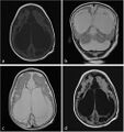
English: Fig. 2. T1-weighted axial (A), T2-weighted coronal (B), T2-weighted axial (C), and axial fluid-attenuated inversion recovery sequence (D) of magnetic resonance images show ventriculomegaly, cystic encephalomalacia, and extensive subcortical and periventricular white matter loss and hyperintensity in white matter with atrophy.
Date
Source https://www.sciencedirect.com/science/article/pii/S1930043317306222
Author Mehmet Sedat Durmaz , Bora Özbakır

Licensing

English: This file is licensed CC BY-NC-ND 4.0

This file was uploaded with UploadWizard.

File history

Click on a date/time to view the file as it appeared at that time.

Date/TimeThumbnailDimensionsUserComment
current21:55, 19 April 2023Thumbnail for version as of 21:55, 19 April 2023470 × 500 (67 KB)Ozzie10aaaaUploaded a work by Mehmet Sedat Durmaz , Bora Özbakır from https://www.sciencedirect.com/science/article/pii/S1930043317306222 with UploadWizard

The following page uses this file:

Metadata